
Introduction
Stomach contraction motility is mediated by a slow wave which is actually an electrical wave generated by the interstitial cells of Cajal (ICCs) in the gastric smooth muscle layer [1-3]. In previous investigations, the alteration of gastrointestinal motility was founded in obese human patients [3,4]. By the same token, it is noted that in rat experiments, intestinal motility regulated by enteric nerve system was shown to be stronger in the evaluated obese group than in the control group [5].
It is noted that medetomidine is an α2-adrenergic agonist drug for analgesia and sedation, which is primarily used in dogs and cats [6-8]. The pharmacologic effects of medetomidine suppress gastrointestinal tract as it inhibits gastric secretion, reticulo ruminal contractions and colonic motility in ruminant and horse, and it is also known to inhibit electrical activity of the small intestine in dogs [6-8]. Additionally, the central nervous system and endocrine functions suppression, as well as muscle relaxation are also included in medetomidine pharmacologic effect [7,8].
The 2-dimensional (2D) speckle tracking imaging is a recent technique for use to evaluate myocardial motion and function assessment in humans [9,10] and in dogs [11-14]. In this way, the 2D speckle tracking traces over a period of time the motion pattern of unique speckle in tissue B-mode images, and is free from the angle parameters, tissue translation, and tethering in the subject [9,11]. In previous studies, speckle tracking and strain measurement were used to assess the activity of an in vitro porcine antrum, in vitro human uterine, and human stomach antral contraction. These evaluations are suitable for a comprehensive quantitative analysis of gastrointestinal and uterine motility in a subject [15-19].
The aim of this study was designed to generate reference data for stomach antral contraction strain by using 2D speckle tracking in dogs, to determine a correlation between the measured variables by the presence of the condition of obesity, and to compare the measured differences between the normal and medetomidine group of subjects in this research study.
Materials and Methods
Animal recruitment
All procedures were approved by the Institutional Animal Care and Use Committee at Gyeongsang National University and the dogs were cared for according to the Guidelines for Animal Experiments (GNU-161031-D0062) of Gyeongsang National University. Clinically 50 healthy dogs were recruited for examination in this study. They were noted to have included 3 intact males, 26 castrated males, 8 intact females and 13 spayed females. The dogs ranged in ages from 1 to 18 years, and were an average weight of 8.1 ± 6.3 kg (range, 2.1 to 37 kg). The dogs were screened for evidence of a gastrointestinal disease by the use of a physical examination, complete blood counts, serum biochemistry, abdominal radiographs, and ultrasonography.
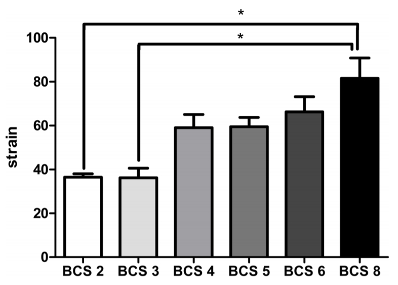
To assess the effect of obesity on stomach antral contraction strain, the body condition score (BCS) was used. It is noted that BCS was commonly used for weight management studies [20]. All dogs were graded and grouped by the BCS score following a physical examination. BCS groups were BCS 2 (n = 3), BCS 3 (n = 5), BCS 4 (n = 10), BCS 5 (n = 20), BCS 6 (n = 9), and BCS 8 (n = 3).
Medetomidine group
Among the 50 dogs, 10 young adult healthy beagle dogs were recruited for the medetomidine group. This group included ten castrated male dogs, and they were all the same age at 4 years old, whereby their average weight was 10.2 ± 1.19 kg (range, 8.6 to 12.2). They had been subjected to a prior ultrasonography examination before the onset of the drug injection. In this case, medetomidine (Domitor®; Orion Pharma, Orionintie, Finland) was injected intravenously into the dogs, with 40 mcg/kg that caused the sedation and analgesia result in all subjects. Next, an abdominal ultrasonographic examination was performed after 5 min after the effect of the drug injection was concluded.
Equipment and measurement
All ultrasonographic examinations were performed by 1 investigator (P.J.H.) using the ultrasound system (Arietta 70; Hitachi Aloka Medical, Tokyo, Japan) and a high-frequency (12 MHz) linear-array transducer. Afterwards, the hair was clipped, and a transducer was applied to the skin of the dog with the use of coupling gel positioned at the right side of the cranial abdomen. Likewise, the dogs were restrained in a dorsal recumbency position on the examination table. By the same token, all of the dogs were fasted 12 h before the start of the ultrasonographic examination.
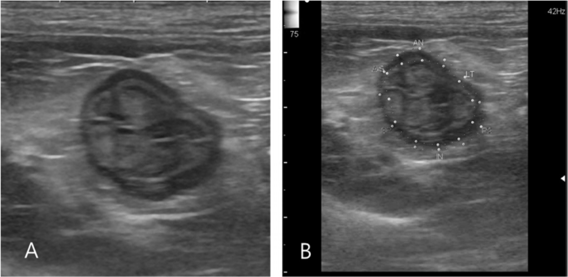
In what follows, the contraction image had been acquired using high quality images possible. Next, the images were analyzed by a same investigator (P.J.H.). Upon review a shot axis view at stomach antrum region was used to evaluate radial strain by 2D-speckel tracking using software program built into ultrasound system. Additionally, all data were obtained from one acquired contraction cycle (Fig. 1A).
In consequence of this study, the resulting strain was therefore mapped in segments of the gastric antrum wall. As noted in the inner and outer borders of the stomach muscular layer, these regions were manually traced to select the appropriate region of interest (ROI) (Fig. 1B). The observer then checked to ensure that the ROI was visually synchronized during the stomach contraction. After processing, the computer software automatically traced the stomach muscular layer and evaluated whether it reliably followed the muscular layer speckles. As an illustration, if the evaluation failed because the software was showed inadequate tracking quality segment, the inner and outer borders were manually corrected and reevaluated. In that case that there were more than three attempts that failed, the entire images were excluded from analysis. The peak systolic strain and the speckle size which were noted at a distance of a two-strain measurement point that formed the basis of the strain estimation in the inner and outer of stomach membrane whereby ROI were measured in the radial plane.
Statistical analysis
All statistical analyses were performed with commercial statistical analysis software (SPSS version 19.0; SPSS Inc., USA). The use of a one-way analysis of variance (ANOVA) was performed to determine whether a relationship existed between antral contraction strain, and BCS in normal dogs. In this case, a one-way ANOVA followed by a post hoc Tukey’s multiple comparisons test was used to compare the peak strain in each of the BCS groups. Next, a paired t-test was performed after normality testing to determine correlations in the antral contraction strain, after administration of the medetomidine in 10 dogs. In the statistical test, a p value less than 0.05 was considered to indicate a significant difference.
Results
Normal range of antral contraction strain and correlation with BCS score
In the entire group of 50 dogs, the mean antral contraction strain was 58.2 ± 20.47% (mean ± SD) and the 95% confidence interval was 52.39% to 64.02%.
Comparison of antral contraction strain between normal group and medetomidine group
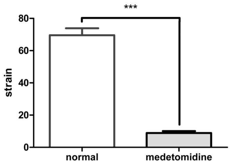
In this case, the mean ± SD (range) of antral contraction strain at normal group was 69.56 ± 13.71% (59.75% to 79.37%), and the medetomidine group was 8.9 ± 3.73% (6.24% to 11.57%). The strain in the medetomidine group was significantly lower than was seen in the normal group (p < 0.001) (Fig. 3).
Discussion
In previous studies, it was shown that the strain measurement in a human antral contraction was 82%, and it also identified that measurement of an intragastric balloon pressure was seen to significantly influence the relationship with strain, therefore strain had a significant correlation with the instance of a stomach contraction [15,17]. In the present study, it is noted that the antral contraction strain is 58.2 ± 20.47% (mean ± SD) in dogs.
Specifically, the use of in vitro testing using a silicon strip phantom mimicking slowly moving tissue was revealed, and that if the speckle size had been small, the strain result was underestimated. Increasing the speckle size to 1.9 mm might reduce some of this error. But speckle size up to 3.1 and 4.2 mm did not increase the accuracy further, and it was shown that the speckle size to 0.8 mm was too small for measuring the strain results for the in vitro phantom experiment [21]. It must be remembered that it was revealed that another study which measured the porcine antral contraction strain had compared 1.2 mm with 1.9 mm speckle size, and it was seen to be better in 1.9 mm speckle size to estimate the strain in in vitro model [16].
In this study, the speckle size was 1.36 ± 0.35 mm (mean ± SD) in dogs. Another key point is that the speckle size was shown to vary individually among the dogs tested. In this case, the testing could not exclude the possibility of an influence to the strain result.
It has been reported that there were many methods to evaluation the stomach motility [22-24]. Among these methods, electrogastrography (EGG) was a traditional noninvasive technique which is often used when evaluating the gastric electrical activity [22-24]. In a human study, children tested by EGG allowed clinical researchers to evaluate the difference in normal and obese patients, as the results were able to reveal that there was no differences found in the normal and obese patients evaluated [25]. But other studies revealed that in morbid obese adult patients, the results also showed an increase in the percentage of bradygastria [26]. All in all, it is noted that bradygastria was associated with a strong antral contraction [27]. Alternatively, it is noted that in the EGG method, there was limitation in measuring obese patients, because the distance between the electrode and the stomach was too far a distance in morbid obesity patients, whereby it could decrease the signal detection and might show the erroneous result for that reason [3]. In this study, the BCS 2, BCS 3 groups showed significant correlation with the BCS 8 group (p = 0.042 and p = 0.016, respectively). It was revealed that the BCS 8 group showed a high strain result, which was higher when compared to the BCS 2 and BCS 3 groups. This result meant that the obese group had a strong antral contractility, as compared with the lean groups.
Medetomidine was revealed to reduce the gastrin secretion in the stomach through an activation of both the central and peripheral α2-adrenoceptors, and it was also revealed that it inhibited the migrating myoelectric complex pattern of the small intestine [6]. In other reports, gastrin secretion inhibition was associated decreasing the motility of the gastric antrum, duodenum, mid jejunum and ileum in dogs [6,28].
In this study, the stomach antrum motility was evaluated by the strain. In this study, there were noted comparisons in the normal and medetomidine groups, whereas the medetomidine group was noted to have revealed a significantly low strain result. This evaluation meant that the stomach motility was found to be decreased in the medetomidine group. Namely, it was the same result as previously reported in dogs [28] that medetomidine effect was found to reduce the gastrointestinal motility.
This study has several limitations. First, the stomach gas revealed disturbed imaging of the entire cross section, as was seen during the ultrasonography examination. This result made it difficult to determine the ROI region in 2D-speckle tracking. The reverberation artifact and shadowed area in 2D-speckle tracking imaging could produce an error in the result [11]. Therefore, the evaluation may have an effect on the strain result for this reason. Second limitation was that excessive adipose tissue also could impair image quality in general ultrasonography imaging [15,17]. Third, in BCS groups there were significant differences in the number of dogs between the groups. These differences may influence the strain results. Further studies are necessary to provide a review of and evaluation of these correlations.
In conclusion, the usefulness of 2D-speckle tracking was verified to measure the antral contraction in dogs. The mean antral contraction strain was 58.2 ± 20.47% (mean ± SD) and the 95% confidence interval was 52.39% to 64.02%. Notably, this is the first report to measure antral contraction strain using 2D-speckle tracking in dogs.
The obesity which was assessed by a BCS score affected antral contraction strain, and it was noted that the obese group showed a strong antral contraction strain than the lean group. Medetomidine also affected the stomach motility and showed the incidence of a low antral contraction strain than was noted in the normal group.
2D-speckle tracking may be useful to evaluation in stomach motility disorders through the antral contraction strain result.
 PDF Links
 PubReader
 Full text via DOI
 Download Citation
 Print
Print


