

Tumors of the penis and prepuce are among the most common neoplasms in Equus caballus species [1]. Penile neoplasms comprise penile papilloma, squamous cell carcinoma (SCC), melanoma, sarcoid, fibrosarcoma and fibroma [2]. Penile papillomas are the second most frequent tumors occurring over the penis and prepuce of horses, and similar to SCC, most develop on the glans penis [3,4]. Penile papillomas are considered as precancerous lesions which can progress to carcinomas in situ and ultimately SCC [3,5]. SCC in the penis is locally invasive, often induce discomfort and can cause serious and even fatal complication [6]. The recurrence of penile SCC is quite high after treatment, and the prognosis of penile SCC is regarded as poor generally [7,8].
Equus caballus papillomavirus type 2 (EcPV-2) is regarded the most clinically [5,7,9], and the association of EcPV-2 and genital neoplasia, including penile papilloma, carcinoma in situ and SCC, in horses has been reported frequently [10-12]. Although penile neoplasm is thought to be common in male horses, however, penile neoplasm affected by EcPV-2 infection have not been reported in Asia yet.
This report provides a pathological description of equine penile neoplasm in a miniature Appaloosa and phylogenetic analysis of EcPV-2 identified from clinical penile neoplasm firstly in Korea.
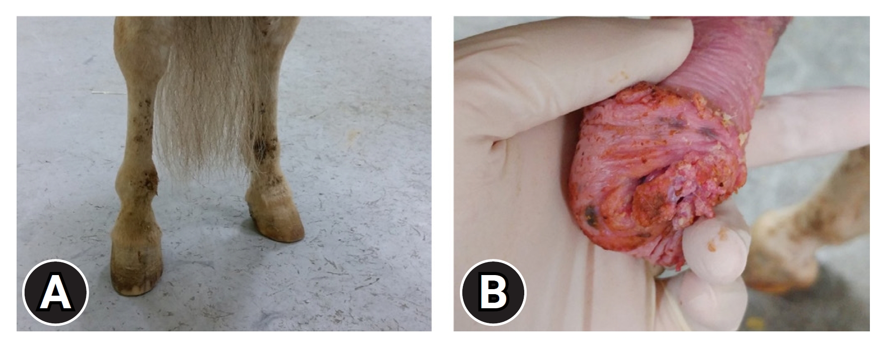
An 18-year-old miniature Appaloosa stallion has approximately 6 months of history of intermittent sanguineous discharge crusts on the inside of hind limbs (Fig. 1A) and recent discomfort of micturition presented to the veterinary center of Korea Racing Authority. No abnormality was found in the hind limbs and ventral abdomen, then multiple ulcerative cauliflower-like masses with diffuse small masses around glans penis were found from the exposed penis after sedation with detomidine (0.01 mg/kg, intravenous [IV], Detomidin; Turkey) (Fig. 1B). Mainly 30 ├Ś 10 ├Ś 11 mm and 10 ├Ś 15 ├Ś 11 mm of 2 cauliflower-like masses around urethral fossa and multiple small masses were presented on glans of penis. Hematologic and serum biochemistry test results were in normal range. Considering small size of tumors and age of the horse, cryotherapy was applied to the horse with notification of recurrence risk to the horse owner. During examination on the penis, the mass was excised. A half of excised main tumor tissue was fixed in 10% formalin solution then submitted to a commercial veterinary laboratory for histopathologic diagnosis. Another half of resected mass was used for EcPV-2 polymerase chain reaction (PCR) test. Six sequence of cryotherapy through a cryoprobe (Histofreezer portable cryosurgical system; Cryoconcepts, USA) with 3 to 4 weeks intervals were performed to remove regrown penile neoplasms around urethral fossa on standing sedation with a combination of acepromazine (0.02 mg/kg, IV, Sedaject injection; Samwoomedian, Korea) and xylazine (0.02 mg/kg, IV, Rompun injection; Bayer Korea, Korea). Throughout the serial cryotherapy, the mass was removed. Recurrence of the tumors were observed 6 months after the cryotherapy. Invasive surgical treatments, including partial phallectomy and sheath ablation or en bloc penile resection and retroversion, were suggested to prevent further recurrence of penile neoplasms, however, the owner refuse because of the old age of the horse, pain, and the possibility of recurrence after the invasive surgery. Under general anesthesia, surgical laser excision of penile masses were performed to reduce hemorrhage, swelling and pain using a diode laser instrument (Multicure; Won Tech Co., Ltd., Korea). Topical treatment of 5% 5-fluorouracil (5-FU) cream (JW 5-FU; JW Pharmaceutical Co., Korea) was applied for 2 to 3 mm size of multiple tumors on the penis 2 weeks after the local laser surgery. 5-FU chemotherapy were performed for 5 months with 2 to 3 weeks intervals. It was revealed via follow-up that the horse was euthanized 4 months later due to the regrowth of the penile neoplasm, pain and other discomfort.
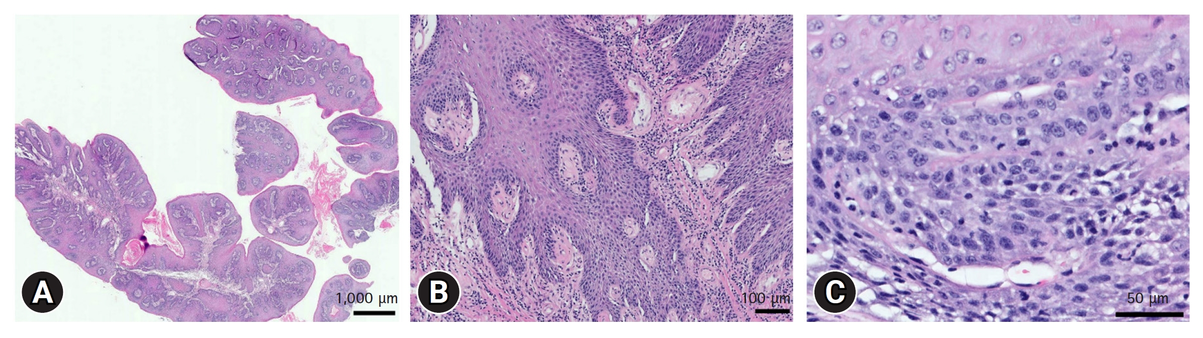
On histopathologic examination, exophytic, papilliferous, poorly demarcated, partially infiltrative neoplasm was composed of polygonal to pleomorphic squamous epithelial cells which were supported by fine fibrovascular stroma (Fig. 2A). The basement membrane was irregular and elongated. Neoplastic cells had a moderate amount of eosinophilic cytoplasm, distinct cell borders, and prominent intercellular bridges (Fig. 2B). Nuclei were round to oval with finely to coarsely stippled chromatin and 1 to 2 distinct nucleoli (Fig. 2C). Mitotic count was 5 per 10 high power fields. Mild anisocytosis and anisokaryosis were observed. The penile mass was diagnosed as papilloma with cellular atypia.
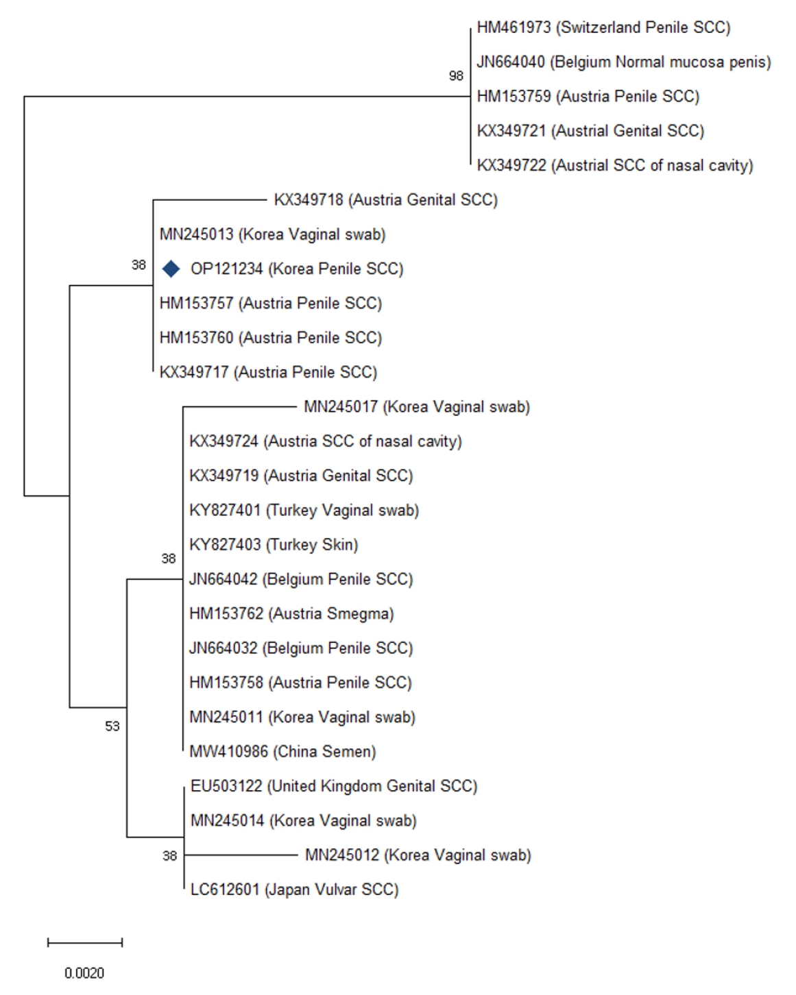
Detection of EcPV2 DNA in the excised mass was performed by PCR targeting EcPV-2 E6 gene [13]. Amplified PCR product containing EcPV-2 E6 gene was sequenced at a commercial sequencing laboratory and compared with previous EcPV-2 sequences using the basic local alignment search tool (BLAST; https://blast.ncbi.nlm.nih.gov/Blast.cgi). The confirmed EcPV-2 E6 gene sequence in this report was submitted to GenBank (OP121234). Phylogenetic analysis of the identified EcPV-2 sequence was performed to evaluate their genetic relationship with 25 sequences from other countries deposited in GenBank using the neighbor-joining method with 1,000 bootstrap values using MEGA 11 software (https://www.megasoftware.net) (Fig. 3). The alignment of the EcPV-2 E6 gene sequences in this study along with EcPV-2 sequences retrieved from GenBank consisted of 327 positions, with nucleotide identities ranging from 98.04 to 99.99%. Korean strain in this study was a match to the retrieved sequences from 3 penile SCCs in Austria and a vaginal swab in Korea (Fig. 3).
In this case, the excised mass of penile neoplasm in a miniature Appaloosa was diagnosed as penile papilloma with cellular atypia, by histopathologic examination with the possibility of carcinoma in situ due to elongated and irregular basal layer of epidermis. Because the characteristics of SCC were observed, including mitotic activity, coarse chromatin, and round or ovoid nucleoli [14]. The mass has nonhealing erosion and cauliflower-like appearance, which are characteristics of penile SCC [2]. Therefore, the penile papilloma treatment approach was treated according to penile SCC treatment method, considering the possibility of being and/or mixed with penile carcinoma in situ.
According to the standardized approach for treating penile tumor [15], radical surgical intervention, such as partial phallectomy and sheath ablation or en bloc penile resection and retroversion, was indicated. However, the owner refused these invasive surgical treatments, considering the old age of the horse, postoperative complications and pain, and risk of recurrence. Less invasive treatments including cryotherapy, local excision, and topical chemotherapy were allowed in this case, despite of the higher recurrence of the neoplasm due to incomplete removal. The reported recurrence rate of penile SCC after local excision or excision combined with cryotherapy appears to be considerably high up to 50% [1]. Equine penile papilloma can progress to carcinoma in situ and eventually to SCC [3-5]. Since only less invasive treatments were applied for prolonged period, it is thought that the complete removal of the penile neoplasm was not achieved through less invasive treatments subsequently the regrowth of the tumor and potentially cancerous progression occurred in this case.
The reported mean age of penile papilloma and SCC were ranged from 16.2 to 18 years [2] and 17.4 to 19.5 years in previous studies [1], respectively. Although exact pathogenesis of penile neoplasm is not proven, but a combination of factors, including lack of pigmentation, smegma and EcPV-2, have been proposed [4]. Breeds with nonpigmentated genitalia such as Appaloosa and American Paint horses, seemed to have a predisposition for penile SCC [1]. Multiple studies have revealed that EcPV-2 infection is etiologically associated with the development of penile neoplasm in horses [4,11,12]. Since the patient was an old (18 years) Appaloosa stallion and EcPV-2 was infected in the penis, penile neoplasm has arisen in the patient in this case.
This is the first molecular detection of EcPV-2 from clinical penile neoplasm in Asia. EcPV-2 have been found in vulvar SCC in Japan [10] and in vulvar swabs in Korea [7]. Penile SCC has been reported in Taiwan [8], but failed the detection EcPV-2 infection. The isolated EcPV-2 E6 gene was compared with 25 sequences from other countries, including Austria, Belgium, China, Japan, Turkey, Switzerland and UK. In phylogenetic analysis, this Korean strain was clustered with Austrian strains from penile SCC and a Korean strain from vaginal swab. EcPV-2 is transmitted through sexual or close-contact between horses [9]. The patient was imported from Australia 6 years before clinical sign onset and has not been used for breeding nor reared with Thoroughbred horses in Korea according to the horse ownerŌĆÖs information; thus, it could not be determined if EcPV-2 in the patient originated from other countries or Korea.
This is the first report of clinical penile neoplasm with EcPV-2 infection in an 18 years old miniature Appaloosa in Asia. But recurrence occurred due to the possibly cancerous progression of the penile neoplasm and the limitation of incomplete removal through less invasive treatments despite of for prolonged period. Thus, early detection and by regular examination of the external genitalia for old male horses with unpigmented genitalia would be beneficial to avoid development and cancerous progression of penile neoplasm, which require radical invasive surgical treatment and tend to recur.
 PDF Links
 PubReader
 ePub Link
 Full text via DOI
 Download Citation
 Print
Print


